conventional X-ray
"X-rays" (German: Röntgen) were discovered by the physicist Wilhelm Conrad Röntgen. X-rays are being ionizised for the purpose of diagnosis in medicine.
For the creation of an image, a short, instantaneous radiation will be emitted. These rays will pass through the body, will
hit an electronic detector behind the body and will be read out there. Based on this information a digital image will be generated
and examined by the radiologist.
The tissues of the human body with different densities absorb the X-rays to different degrees, so that an image of the inside
of the body will be obtained (shading, brightening and other X-ray signals).
Basically every part of the body can be X-rayed. Most frequently the lungs, abdomen and bones, in particular the special orthopaedic areas are imaged. The procedure is often
used, for example, when a bone fracture is suspected: If the X-ray shows an interruption of the continuity of the bone, the
suspicion is confirmed. If kidney stones or gallstones are suspected, an X-ray may also be useful. In some cases, a contrast
agent can be injected into a vein.
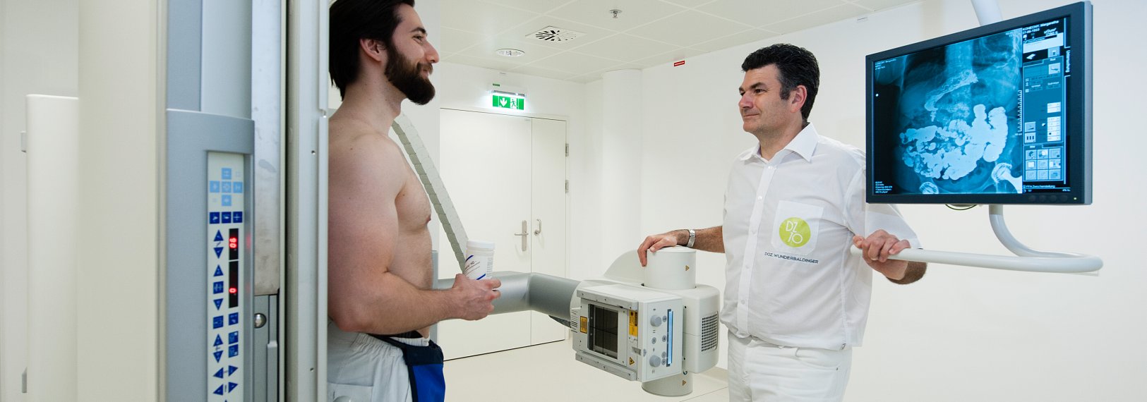
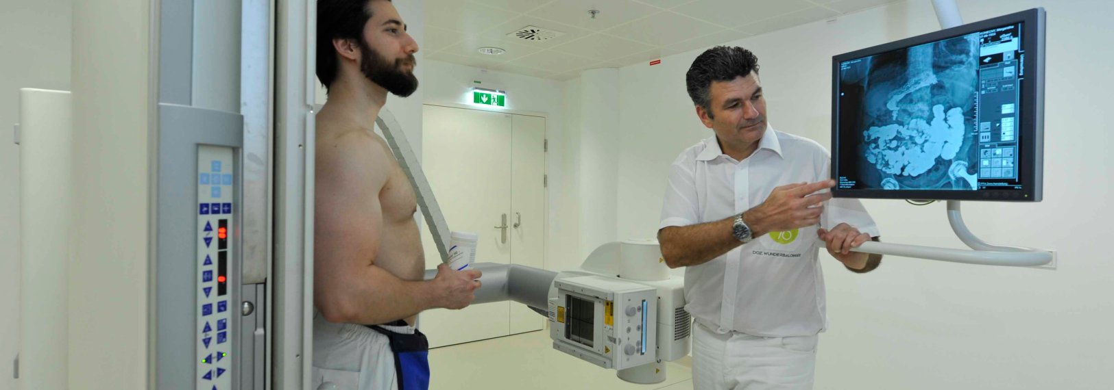
While conventional X-rays only produce one standing image at a time, fluoroscopy takes several sequential images. Up to 15 frames per second are produced here. These are transmitted directly to a monitor and can be viewed like a video film.
The modern devices are additionally equipped with movable tilting tables, which allow an examination in different body positions. It is very important that the patient helps us when we perform a
fluoroscopy with his focus and active compliance.
In addition, contrast agents are often used in these examinations. These are used as needed depending on the examination region and technique. There are
contrast agents which are orally taken as a liquid or introduced through an intestinal tube, other contrast media are injected
directly into the vein.
information on the individual examinations can be found here.
The examination is used to image the pharynx, larynx, oesophagus and stomach. These regions can be investigated dynamically and functional disorders can be identified. The doctor can also detect morphological
changes such as diverticula, scars and tumors.
During the examination, the transport of a swallowed contrast agent through the esophagus will be filmed and evaluated afterwards. It may be possible to take in a paste or even other food mixed with a contrast agent
in addition to normal liquid contrast agents.
If you swallow up often, let the doctor know before the examination so that he can choose the appropriate contrast agent for
the examination.
Gastric X-rays are used to visualize the stomach and duodenum after oral administration of a contrast agent and effervescent powder. When
your stomach is x-rayed, it is extremely important for you to comply with the preparation steps: Don't eat, drink nor take tablets for 3 hours before the examination and don't smoke within that time frame. Important medication can be taken up to 3 hours before the examination with a sip of water. The examination should therefore
ideally take place early in the morning.
Before the colon examination (irrigoscopy), the colon must first be completely cleansed. This is very important, as otherwise possible residual stool could be misinterpreted
as a tumor. The colon has to be completely cleansed before this examination, this is very important. Check out here how to
use laxatives (link).
After the insertion of an intestinal tube, a barium-containing contrast agent and air are delivered into the large intestine.
The patient will then be moved into different positions so that the contrast medium can spread well. The better the contrast
agent will be distributed, the better it will condensate to the wall of the colon and the easier the images can be analyzed
by the doctor.
Optionally, a medication can be injected (Buscopan), which reduces the movement of the intestine, so that a good dilatation
of the intestinal loops will be achieved. However, this medicine may decrease your ability to drive. Whenever your colon is
going to be checked, it would be advisable for you not to drive to the hospital, but rather to be dropped off.
The patient is brought into a tilted, but fixed position of the table on which he is laying. After puncturing a vein on the
back of the foot, a contrast agent containing iodine is injected into the patient. If you are allergic to an iodine-containing
contrast agent, please let the doctor know. Further information on contrast agent allergies can be found here (link).
A feeling of warmth can occur when the contrast agent is injected. The veins are then analyzed while the fluoroscopy is being
performed. Also, the venous drainage can often be blocked first in order to keep the contrast medium in the examination area
for a while.
The purpose of this examination is to visualize the urinary tract. Approximately 50 ml of an iodine-containing contrast agent will be injected into an arm vein for the examination. This contrast
agent will pass the kidneys afterwards. It can be checked whether there is an obstacle to drainage in the area of the urinary
tract. X-rays of the abdomen are taken here at regular intervals (every 15 - 30 minutes). Often, additional images will be
taken after micturition.
All diagnostic X-ray examinations will be performed within a safe dose range. Even multiple examinations in the course of the year are considered harmless. An individual risk evaluation - depending
on the region examined and the medical necessity - is required in the case of a high accumulation (if X-rays are taken several
times a month over a period of years).
All diagnostic X-ray examinations will be performed within a safe dose range. Even multiple, repetitive examinations in the course of the year are considered harmless.
X-rays will not be stored in the human body. They will pass the body and will be distracted and weakened.
In general, ionising radiation can cause genetic changes. However, since the body can repair such changes, radiation rarely
has side effects. These effects can only occur in cases of high-dose radiations. However, our examinations will never reach
this threshold.
No.
Please let our team always know whether you are pregnant.
If pregnancy is possible, please let us perform the examination when a pregnancy test has been negative or at least within
a range of ten days after your most recent period.
Although many images are taken one after the other, the radiation dose can be kept low by using "pulsed" X-rays. Here, every
image is taken only with a small dose of radiation. In addition, modern apertures and pre-filters as well as the principle of "last image hold" ensure a further reduction of
the radiation dose.


