Magnetic resonance imaging (MRi)
In magnetic resonance imaging, patients are placed in a magnetic field. This stimulates the hydrogen molecules in the body. By means of this signal sectional
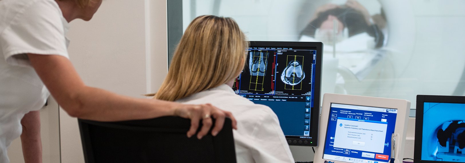
images can be generated which, in addition to the anatomy of the body, can also show inflammatory changes and other pathologies with very high accuracy. The images are sent directly to a computer for evaluation.
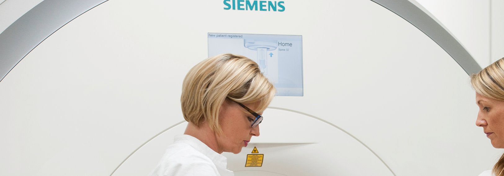
With the newly installed 3 Tesla magnetic resonance tomograph, the Skyra MRT from Siemens, a high-end device is available to our patients. This device offers an extremely high resolution and shorter examination times.
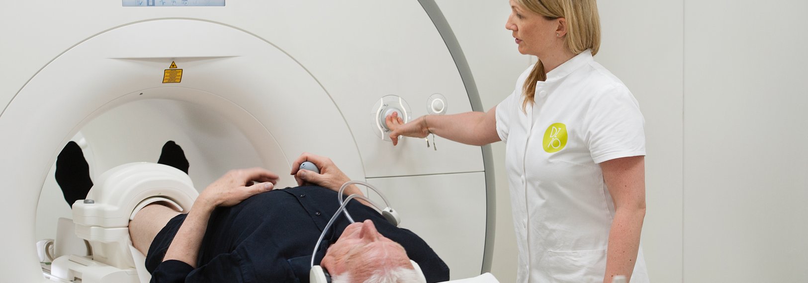
The duration of the examination is 15 to 30 minutes. During this time you will have an alarm bell in your hand so that you can communicate with the assistant at any time. For the evaluation of the examination, it is important that you
don´t move.
Overview of all examinations
Skull:
Standard (brain and facial skull)
Diffusion imaging
Brain nerves:
Jaw joints (incl. dynamics)
MR angiography of intracranial vessels
Neck:
Standard (soft tissues, salivary glands)
MR Sialography
MR angiography of extracranial vessels
Chest (Thorax):
Standard (Mediastinum)
MR mammography with contrast medium dynamics
MRI of the heart
MR angiography of the aorta and pulmonary arteries
Abdomen:
Standard (liver, pancreas - pancreas, spleen, kidneys and adrenal glands - retroperitoneum, lymph nodes)
MRCP (gall bladder and ducts)
MR enteroclysis
MR angiography (aorta, renal arteries, etc.)
MR urography
Pelvis:
Standard (uterus, ovaries, endometriosis)
Fistula MR
Prostate (multiparametric evaluation, PIRADS):
80% of the examination fees are now reimbursed by the health insurances. To this end, a chief physician (a specialist urology
doctor) has to approve this in writing.
You will receive an invoice from us at the institute, which you can then submit to your health insurance company.
Joints/Bones:
Standard (all bones and joints, entire spine including intervertebral discs, bone marrow)
MR arthrography
cartilage diagnostics
Diffusion Imaging (Spine)
Myelography
Other examinations:
Pelvis/leg MR angiography
Full body MRI
special examinations
MR Enteroklysma
Preparation for the special examination MR enteroclysis:
- Diet
- Cleanse your colon by means of drinking liquids
Diet
Foods that leave indigestible residues should be avoided on the day before the examination. These are e. g. fibres of meat,
vegetables, fruits and nuts. Also dairy products, as for example cheese, can only be slowly digested. Please don´t eat any
bread.
CLEANSE YOUR COLON BY MEANS OF DRINKING LIQUIDS
In addition to dietary preparation, cleansing and washing out the intestines is extremely important. Please drink as much
liquid as possible (3-4 litres per day) during the preparation period. You can drink liquids also on the day of your examination.
However, please comply with the doctor´s recommendation about the amount of liquids on that day. schedule
- Day before examination: Diet (see above)
- Day of examination: Don´t eat, only drink.
- 2 hours before the examination, you will be given a special drink to mark the intestine.
Multiparametric MRT of the prostatic gland with PIRADS report:
80% of the examination fees are now reimbursed by the health insurances. To this end, a chief physician (a specialist urology
doctor) has to approve this in writing.You will receive an invoice from us at the institute, which you can then submit to your health insurance company.
Patients who are referred by a physician who is paid by the obligatory insurance do not need to obtain a referral approved
by a chief physician. If referrals are issued by physicians of your choice, a chief physician's permit is required. This permit
has to be obtained ahead of the examination. Please always bring your ECARD.
2-3 hours before the examination you should not eat anything.
During the examination (depending on the region of examination and/or indication) the doctor may inject you with contrast
medium via a venous access.
As the examination requires your written consent, we ask you to come 20 minutes before the scheduled appointment to fill in
the consent form and a general questionnaire about your medical history.
Patients with prostheses, implants after surgeries, such as screws and plates, hearing aids, pacemakers, heart valves, stents (these are small metal tubes in vessels, usually
coronary vessels) have to report above-mentioned items before the MRI examination, since magnetic fields are used in MRI for
imaging. Please bring your implant ID - if available.
Jewellery, hairpins, glasses, hearing aids and coins must be placed in the changing room if possible.
Non-removable metal parts that are in the body, such as metal splinters and certain red dyes used in tattoos and permanent
eyelid strokes, may contain iron oxide and therefore warm up or move during the examination and must therefore be reported before the examination.
We are going to decide this on a case-by-case-basis. Therefore, it may also happen that a doctor might inject you a contrast agent in the course of the examination.
A pregnancy is not necessarily a contraindication to an MRI, but due to the lack of data an examination in the first third
of a pregnancy is not performed. Please report when you are pregnant. Then, we won't inject any contrast agents.


